Smooth Muscle Diagram : Smooth Muscle Diagram - The Sources Of Synthetic Vascular ... : The gi tract stretches from the mouth to the anus.. Smooth muscle (factors affecting activation, general properties, source of cytosolic ca2+, structure, muscle cells). Smooth muscle (factors affecting activation (spontaneous electrical…: Smooth muscles are mainly divided into two subgroups: Smooth muscle and cardiac muscle move to facilitate body functions like heartbeats and digestion. By ning zhou, shaunrick stoll, christiana leimena and hongyu qiu.
Smooth muscle is found in the walls of hollow organs like your intestines and stomach. It constitutes much of the musculature of. Neuromuscular junction vector illustration scheme. Smooth muscle and cardiac muscle move to facilitate body functions like heartbeats and digestion. Because visceral muscle is controlled by the unconscious part of the brain, it is known as involuntary muscle—it cannot be directly controlled by.
Muscle Tissue Etc. - Physio with Berry at Sir Francis ... from classconnection.s3.amazonaws.com Cardiac muscle vector illustration diagram, anatomical scheme with human heart. The gi tract stretches from the mouth to the anus. The trichome stain can be used to highlight smooth muscle cells (red) and background collagen (blue) in cases of spindled cell tumors. Vascular smooth muscle cells (vsmcs) are the stromal cells of the vascular wall and are responsible for regulating arterial tone, blood pressure, and blood supply of the tissues. Smooth muscle contraction requires both myosin activation and actin cytoskeletal remodeling. By ning zhou, shaunrick stoll, christiana leimena and hongyu qiu. In suvsm, a single smooth muscle cell in a bundle is innervated by an autonomic nerve fiber. The term smooth muscle refers to a muscle of the human body that is part of an involuntary muscle group.
Circuit diagram used for study 1141x1080 draw the diagram of smooth muscles or neuron muscle
It is the weakest type of muscle but smooth muscles in the gastrointestinal or gi tract control digestion. By ning zhou, shaunrick stoll, christiana leimena and hongyu qiu. The term smooth muscle refers to a muscle of the human body that is part of an involuntary muscle group. The image above shows how the actin and myosin fibers shorten, effectively shrinking the cell. Smooth muscle (factors affecting activation, general properties, source of cytosolic ca2+, structure, muscle cells). Although smooth muscle is located in many different parts of your body, this session focuses on the smooth muscle that is located in the intestine. *smooth muscle* the cardiovascular, gastrointestinal, genitourinary, and respiratory systems are composed mostly of hollow organs (tubular or sacular) other structures in the body that contain smooth muscle include the myometrium — the muscular wall of the uterus — which is responsible. Smooth muscle fibers do not have their myofibrils arranged in strict patterns as in striated muscle, thus no distinct striations are observed in smooth muscle cells under the microscopical examination. Smooth muscle structure, embryonic origin, and histology. 12 photos of the smooth muscle diagram. Vascular smooth muscle is the type of smooth muscle that makes up most of the walls of blood vessels. Like all muscle tissue, the function of smooth muscle is to contract. In this video i have shown the simplest way of drawing muscle drawing.
You will have some basic understanding of the appearance referring to the below smooth muscle diagram. Diagram of systems in human body. *smooth muscle* the cardiovascular, gastrointestinal, genitourinary, and respiratory systems are composed mostly of hollow organs (tubular or sacular) other structures in the body that contain smooth muscle include the myometrium — the muscular wall of the uterus — which is responsible. Attached to the bones of the skeletal system are about 700. In suvsm, a single smooth muscle cell in a bundle is innervated by an autonomic nerve fiber.
The Differences Between Skeletal, Smooth & Cardiac Muscles ... from usercontent2.hubstatic.com The trichome stain can be used to highlight smooth muscle cells (red) and background collagen (blue) in cases of spindled cell tumors. The term smooth muscle refers to a muscle of the human body that is part of an involuntary muscle group. Attached to the bones of the skeletal system are about 700. Learn how your gut contracts! It is divided into two subgroups; Smooth muscle histology and diagram (inlet). You can also find smooth muscle in the walls of passageways, including arteries and veins of de cardiovascular system. Vascular smooth muscle refers to the particular type of smooth muscle found within, and composing the majority of the wall of blood vessels.
Smooth muscle, muscle that shows no cross stripes under microscopic magnification.
It constitutes much of the musculature of. Vascular smooth muscle refers to the particular type of smooth muscle found within, and composing the majority of the wall of blood vessels. Smooth muscle (factors affecting activation, general properties, source of cytosolic ca2+, structure, muscle cells). Smooth muscle, muscle that shows no cross stripes under microscopic magnification. Smooth muscle (factors affecting activation (spontaneous electrical…: Smooth muscle fibers ____x smaller than fibers in skeletal muscle. Circuit diagram used for study 1141x1080 draw the diagram of smooth muscles or neuron muscle Smooth muscle and cardiac muscle move to facilitate body functions like heartbeats and digestion. Smooth muscle has a fusiform shape, which resembles a football or spindle. Vascular smooth muscle cells (vsmcs) are the stromal cells of the vascular wall and are responsible for regulating arterial tone, blood pressure, and blood supply of the tissues. Smooth muscle is also called involuntary muscle or unstriated muscle. Fibers insulated from each other by covering of collagen and glycoprotein fibrillae. The image above shows how the actin and myosin fibers shorten, effectively shrinking the cell.
Visceral muscle tissue, or smooth muscle, is tissue. The gi tract stretches from the mouth to the anus. This is different from cardiac muscle tissue, which develops into an as you look at this diagram of a smooth muscle fiber, you'll notice the single nucleus in the center. Smooth muscle is a type of muscle tissue which is used by various systems to apply pressure to vessels and organs. It is divided into two subgroups;
Muscle tissue - Wikipedia from upload.wikimedia.org Smooth muscle (factors affecting activation, general properties, source of cytosolic ca2+, structure, muscle cells). Muscular system anatomy diagram & function smooth muscle smooth muscle makes up the walls of hollow organs respiratory passageways and blood vessels its wavelike movements propel muscle and cardiac muscle the muscular system simple diagram muscular system unlabeled diagram. Keep reading to learn more about smooth muscle examples and how they function in the body. Smooth muscle and cardiac muscle move to facilitate body functions like heartbeats and digestion. Although smooth muscle is located in many different parts of your body, this session focuses on the smooth muscle that is located in the intestine. Smooth muscle (factors affecting activation (spontaneous electrical…: Smooth muscle contraction requires both myosin activation and actin cytoskeletal remodeling. This is different from cardiac muscle tissue, which develops into an as you look at this diagram of a smooth muscle fiber, you'll notice the single nucleus in the center.
Cardiac muscle vector illustration diagram, anatomical scheme with human heart.
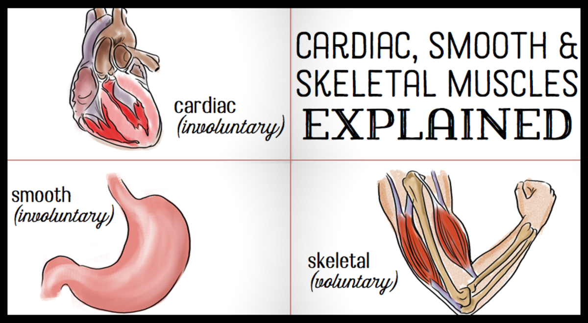
Smooth muscle, muscle that shows no cross stripes under microscopic magnification. In this video i have shown the simplest way of drawing muscle drawing. Keep reading to learn more about smooth muscle examples and how they function in the body. Although smooth muscle is located in many different parts of your body, this session focuses on the smooth muscle that is located in the intestine. Smooth muscle (factors affecting activation (spontaneous electrical…: Diagram of artery with smooth muscle identification. Ciliary muscle of eye, iris, piloerector muscles. Smooth muscle lines the inside of blood vessels and organs, such as the stomach, and is also known as visceral muscle. Diagram of systems in human body. 12 photos of the smooth muscle diagram. Vascular smooth muscle refers to the particular type of smooth muscle found within, and composing the majority of the wall of blood vessels. It is the pen diagram of skeletal, smooth and cardiac muscle for class 10, 11 and 12. You can also find smooth muscle in the walls of passageways, including arteries and veins of de cardiovascular system.


.svg/1920px-Muscle_Tissue_(1).svg.png)


0 Komentar